Radiographic Anatomy
Comprehensive anatomy by body region — bones, joints, muscles, soft tissues, and radiographic landmarks
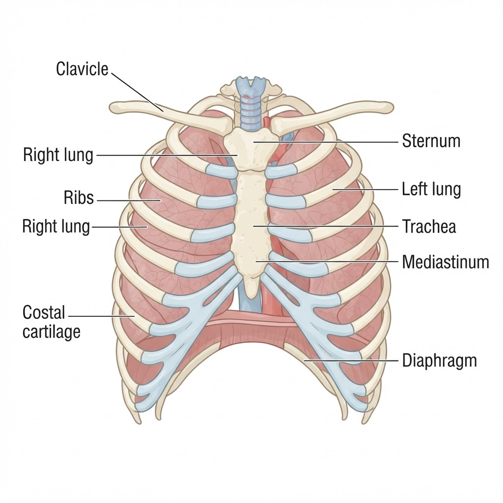
Thoracic Cage — Anterior View
AnteriorRibs, Sternum & Lungs
Anterior view of the thoracic cage demonstrating the sternum, ribs, costal cartilages, clavicles, lungs and mediastinum — the anatomical foundation for chest radiographic positioning.
Thorax
- Sternum: manubrium, body, xiphoid process
- 12 pairs of ribs (7 true, 3 false, 2 floating)
- Thoracic vertebrae T1–T12
- Clavicles (×2)
- Scapulae (×2)
- Sternoclavicular joints
- Costochondral junctions
- Costovertebral joints
- Costotransverse joints
- Manubriosternal joint (Angle of Louis — T4/5 level)
- Diaphragm
- External intercostals
- Internal intercostals
- Pectoralis major
- Pectoralis minor
- Serratus anterior
- Heart (4 chambers — RA, LA, RV, LV)
- Lungs: Right (3 lobes — upper, middle, lower), Left (2 lobes — upper, lower)
- Trachea (bifurcates at T4/5 — carina)
- Main bronchi
- Aorta (ascending, arch, descending)
- Superior and inferior vena cava
- Pulmonary arteries and veins
- Esophagus
- Thymus
- Mediastinum (anterior, middle, posterior)
- Jugular (sternal) notch — T2/3
- Angle of Louis (manubriosternal junction) — T4/5
- Xiphoid process — T9/10
- Inferior angle of scapula — T7
- Vertebra prominens (C7/T1)
PA chest: both lung fields, cardiac silhouette (CT ratio ≤ 0.50), mediastinum, trachea midline, hemidiaphragms, costophrenic angles. Normal cardiac borders: right = SVC + right atrium; left = aortic knob + pulmonary artery + left ventricle. Carina angle normally < 70°.
Understanding this region's anatomy is essential for accurate patient positioning, image evaluation, and pathology identification. Key landmarks guide central ray placement and ensure optimal demonstration of the structures of clinical interest.
No anatomy diagrams available for this region
Anatomy diagrams for this body part will appear here once added.
References Used
Bontrager's Textbook · Merrill's Atlas · Clark's Positioning · Gray's Anatomy · OpenStax Anatomy & Physiology · Radiopaedia · StatPearls · ACR · ESR · RSNA · WHO
All educational content is originally written based on the medical concepts from these trusted references. No copyrighted text, tables, or illustrations are reproduced.